Dermatovenerology investigations and tests

Services

I have a rich case history and a wide clinical experience in the diagnosis, prevention, and management of various dermatological and venereological pathologies in both women and men.

My areas of dermatological medical interest include:

  • - Dermoscopy
  • - Cryotherapy
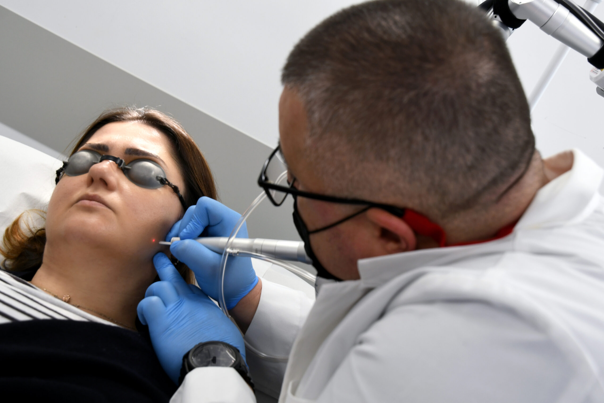
  • - Dermatological laser therapy
  • - Dermatological screening of skin lesions
  • - Prevention of various dermatological conditions

During consultations you will receive a correct diagnosis and appropriate medical treatment for skin conditions from all 24 categories of skin diseases (e.g., psoriasis, seborrheic dermatitis, eczema, rosacea, acne vulgaris, skin atopy / atopic dermatitis, shingles, various skin viruses and bacterial skin infections and skin cancer, etc). Research training and knowledge of medical genetics give me a unique perspective on dermatological conditions from the spectrum of rare diseases and genodermatoses.

A mandatory dermatological consultation for various conditions requires a thorough conversational review, intended to elicit additional data and details (often very valuable for diagnosis) related to the reason for the consultation, as well as an objective clinical examination often facilitated by a dermatoscope or various other equipment optics for dermatological use. Occasionally, additional investigations are needed to document the clinical case. These may include laboratory tests (blood), examination under UV light (Wood's lamp) or taking a small lesional skin sample (lesional biopsy). The diagnosis and therapeutic scheme will emerge as a result of such an investigative process.
Only skin lesions that provide clinical and dermoscopic diagnostic certainty benefit from laser, radiofrequency, or electrocautery removal services. The existence of the smallest point of diagnostic equivocation will require prior lesional biopsy, followed by the dermatopathological examination of the biopsy piece. All aspects of a fair and complete treatment are exposed and discussed beforehand with the patient and carried out only after giving their informed consent.

Dermatological consultation

This represents a meeting between the dermatologist and the patient in the dermatology office, in the context for which the patient applied for skin or mucous membrane problems. During this consultation, a detailed medical history is taken on the patient's health status, including the patient's family medical history, current medication, diet or, in the case of women, pregnancy or breastfeeding status. Then the anamnesis and the objective examination of the patient's skin are performed. Sometimes, depending on the symptoms, additional investigations are necessary (bacteriological, mycological, allergological testing, examination with UV light, biopsy, etc.) to establish the diagnosis with certainty, after which the diagnosis will be decided.

At the time of a final diagnosis, a personalized action plan is created, which will involve medical treatment and/or surgical, laser, or dermato-cosmetic treatment. The patient is always provided with detailed explanations, directions, information and advice regarding the received diagnosis, the treatment, the care to be given at home or if further investigations (sometimes even by other medical specialties) are required. The need for the follow-up will be also assessed and mentioned to the patient.

Wood lamp examination

Examination in UV light with the Wood lamp is an extremely effective diagnostic test for certain types of dermatological conditions (e.g., pityriasis versicolor, tinea capitis, erythrasma, vitiligo and melasma).

Dermoscopy

Dermoscopy is a modern, fast, and non-invasive dermatological investigation method, used to increase the accuracy of the examination of skin lesions. It involves the use of the dermatoscope (a hand-held microscope) applied to the surface of the skin, which provides a magnified image of the lesion to be examined, allowing the observation of skin lesion details, usually hidden from naked eye observation. In general, dermoscopy is used to diagnose pigmented skin lesions (e.g., nevo-cellular nevus, melanoma) or other proliferative lesions, either benign (e.g., seborrheic keratoses) or malignant (e.g., basal cell carcinoma, squamous cell carcinoma), and to detect lesion changes that may indicate a pre-cancerous or cancerous lesion, or any other inflammatory conditions.

Cryotherapy

Cryotherapy, also known as "cold therapy" is a dermatological therapeutic procedure that uses liquid nitrogen applied to the skin for short periods of time, targeted on areas of various sizes. It represents a very effective procedure in the rapid removal (with minimal discomfort) of benign epidermal lesions (e.g., papilloma, seborrheic keratoses, flat warts, actinic keratoses). It does not require anesthesia and can be applied to skin areas with aesthetic involvement. It is relatively easily accepted by children and has very good cosmetic results.

Bio-revitalization therapy

Synonymous with "vampire therapy", bio-revitalization therapy uses platelet rich plasma (PRP). Through this minimally invasive procedure, the growth factors in the skin are supplemented. This procedure is very effective in obtaining a facial lifting effect, reducing expression lines, fine wrinkles, and stimulating hair growth.

Seborrheic keratoses

Seborrheic keratoses are some of the most common dermatological tumor formations. In the case of a definite diagnosis, they can be removed using a CO2laser or radiofrequency ablation (an electro-scalpel that operates in the medium radio frequency band).

Molluscum pendulum

Molluscum pendulum is a small tumor mass with dimensions between 2-10 mm, anchored to the skin of the envelopes by a pedicle of variable sizes. It can be excised by laser and/or radiofrequency and, for diagnostic accuracy, can benefit from dermato-pathological examination.

Papilloma

Cutaneous papilloma is an exophytic, non-cancerous neo-formation lesion with a contagious risk, which represent a reservoir of cells infected with the HPV virus. They require treatment to remove the lesion, thus preventing spread by autoinoculation and/or transmission by direct contact to other people.

Condyloma

Genital condylomas are small exophytic lesions, growing on the surface of the skin of the female and/or male genital organs as a result of infection with the HPV virus (especially strains 6 and 11) through skin-to-skin contact. Screening for sexually transmitted diseases is required before excision.

Warts

Vulgar warts are benign, frequent lesions due to human papillomavirus (HPV) infection. The skin subjected to microtraumas is frequently affected, such as the skin covering the extremities, the skin (but also the mucous membrane) of both genital and oral areas. It is transmitted directly through skin-to-skin contact. Under local injectable or epicutaneous anesthesia, they can be excised by different methods.

Xanthelasmas

Xanthelasmas are small, yellow-brown, soft, asymptomatic plaques located on the eyelids. It represents fat deposits localized in the dermis, which have a slow evolution over the years, especially affecting people in their 50s.

Cutaneous hemangiomas

Cutaneous hemangiomas are sites of abnormal vasculature of the skin, characterized by an increased agglomeration of blood vessels (usually of neo-formation) in a well-defined cutaneous region. The location can be varied on the trunk, upper and lower limbs, paracervical or scalp. There are frequent cases (especially in children) in which they disappear by themselves. Medical consultation is indicated in all situations, therapeutic solutions in many cases involves laser vaporization of these formations.

Molluscum contagiosum

It represents a viral infection of the skin (caused by a poxvirus) characterized by particular lesions, easily identified in the context of a dermatological consultation. Usually, the lesion is featured by an eruptive progression. The mode of transmission is through direct skin-to-skin contact (with transmission from affected skin to undamaged skin), or through contact with various contaminated objects.

Sebaceous adenoma

Sebaceous adenoma is a benign tumor formed by the exuberant development of incompletely matured secretory cells of the sebaceous gland. It appears most of the time as a solitary nodule located predominantly on the face and scalp (but also in other skin regions such as the scrotum), of a yellowish-white color, with a firm consistency. It is prevalent in older people.

Syringoma

These represent benign tumors originating from the cells of the eccrine sweat glands. They are located at the level of the eyelids (especially the lower eyelid), appearing either isolated or with eruptive evolution at the level of the skin of the upper chest (anterior and posterior). They are monomorphic lesions, small papules of 1-3 mm or firm, skin-colored, hemispherical nodules and asymptomatic. Their spontaneous regression is exceptional, usually they are being removed by request.

Epidermoid cyst

It is a benign tumor resulting from the occlusion of the hair follicle, which is why it is most frequently located on hairy skin. It acquires a hemispherical appearance, yellowish-white in color, with either an elastic or firm consistency. One variety is the milium cyst(s), which are yellowish, multiple, punctate, and have a cornified consistency. Their spontaneous regression is exceptional, most of the time they are being removed by patient’s request.

Keloid scars

Keloids are spontaneous or post-traumatic tumors that appear especially in areas with an abundance of soft tissue. The triggering factors induce the proliferation of fibroblasts, which will subsequently cause an increased production of collagen fibers, followed by their densified agglomeration in well-defined sites. This creates reddish hypertrophic lesions, firm with a smooth surface, sometimes accompanied by a local painful sensation. The elimination of these types of injuries is procedural, involving local injections and/or laser therapy.

Dermatofibroma

Dermatofibroma is a benign skin tumor that develops in the dermis, more frequently in women, indicating a certain predisposition to fibroids with other locations (e.g., uterine fibroid). It is usually a solitary, nodular, palpable, pinkish-brown lesion, frequently located on the limbs, occasionally on the trunk. Sometimes it can reach dimensions larger than 1 cm in diameter, at which point it is alarming. Correct excision of dermatofibromas is strictly surgical.

Melanocytic nevi

Melanocytic nevi are circumscribed pigmentary dysembryoplasias of the skin or mucosa, which are produced by the excess accumulation of pigment cells. They can be congenital (present at birth) and/or acquired (appearing during life). Their onset is favored by skin phototypes I and II, exposure to sunlight (UV radiation). The most correct approach for these lesions is consisting of an initial dermoscopic assessment, follow-up, or after necessity, the surgical excision with dermatopathological analysis of the excised tissue piece.

Basocellular carcinoma

Studies indicate basal-cell carcinoma to be the most common human malignant tumor, originating from the basal cells of the epidermis and/or adnexa. It is more common in men, especially after 50 years old, primarily located on sun-exposed skin areas. It involves a differential diagnosis, which is why biopsy of these lesions is common. The most correct approach for these lesions is surgical excision with dermatopathological analysis of the excised tissue piece.

Squamous-cell carcinoma

Squamous-cell carcinoma is a malignant cutaneous tumor starting from the keratinocytes of the epidermal spinous layer. It represents the most common tumor of the mucous membranes and presents a significant risk of metastasis. It frequently appears on skin or mucous areas exposed to the sun. Men are more affected than women. A histopathological examination is mandatory in establishing the diagnosis, and the correct treatment is surgical.

Actinic keratosis

Actinic keratosis is a precancerous skin lesion, being the most common clinical lesion encountered in Caucasians. It is favored by advanced age, predominantly male, light skin phototypes, intense exposure to UV radiation and neoplastic antecedents. Histopathological examination establishes the diagnosis. It may benefit from medical treatment, photodynamic therapy, or surgical management.

Acne scars - fractional CO2 laser

Fractional CO2 laser is used effectively in the treatment of post-acne scars due to its ability to stimulate collagen production. By creating thermal micro-columns in the skin, this type of laser promotes the regeneration of damaged tissue. Results include a visible smoothing of the skin texture and a reduction in the depth of post-acne scars. The procedure often requires multiple sessions and involves a short recovery time.

Melasma of the face - cryotherapy / pharmacological depigmentation / fractional CO2 laser

Melasma is a dermatological condition characterized by the appearance of hyperpigmented spots, usually on the face, more frequently in women. It is influenced by hormonal factors, sun exposure and genetic predisposition. Treatment includes rigorous photoprotection, various formulas of topical depigmenting drugs, but also procedures such as laser or cryotherapy. Results vary depending on the severity and individual response to treatment.

Striae distensae / striae gravidarum (popularly known as "stretch marks") - fractional CO2 laser
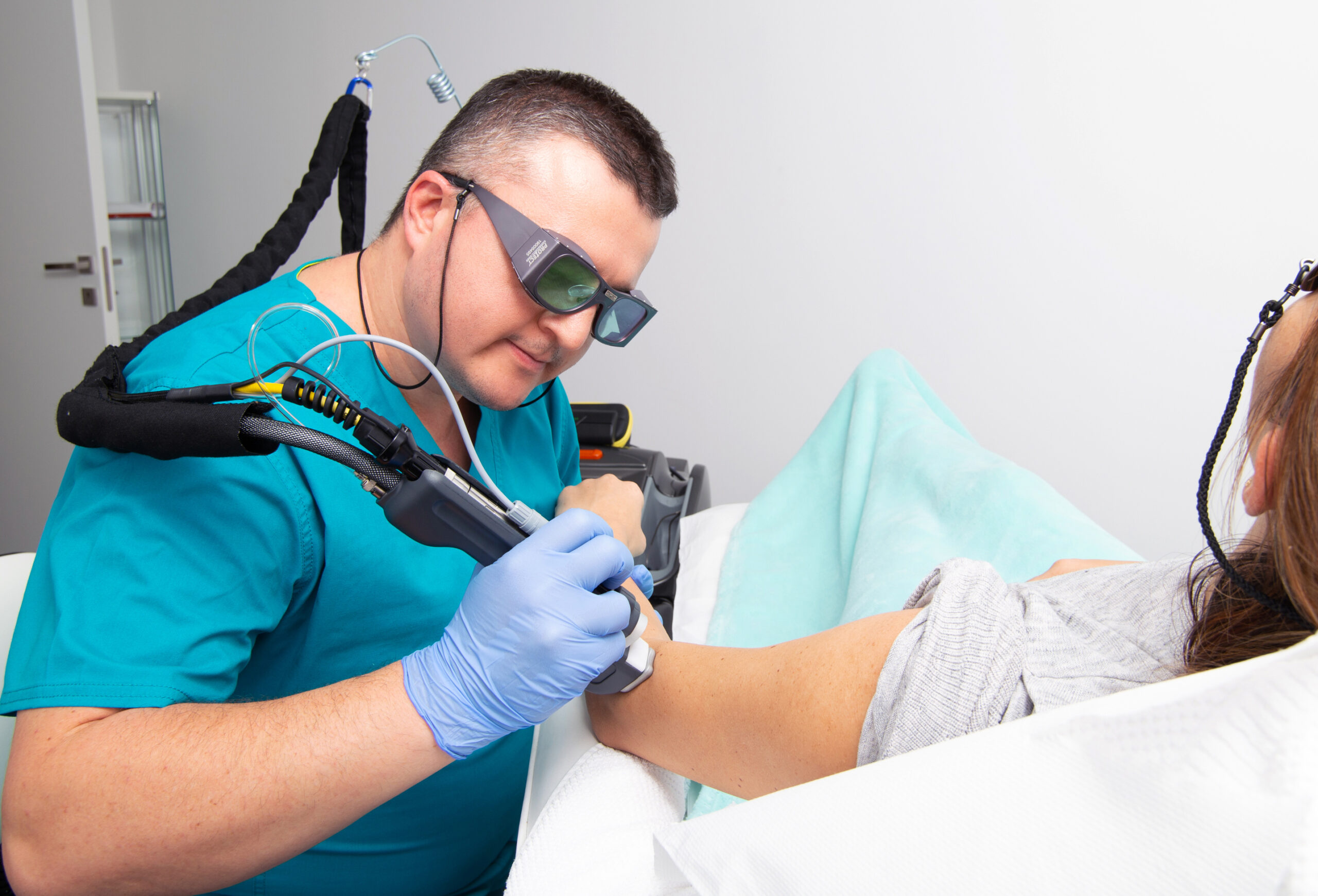
Fractional CO2 laser is successfully used in the treatment of stretch marks (striae distensae), by stimulating the regeneration of collagen and elastin. The procedure creates controlled microlesions in the skin, which favor the restoration of the affected dermal structure. The treatment can improve the texture and color of stretch marks, reducing their visibility. Multiple sessions are required for optimal results, and recovery is usually rapid.

I have a rich case history and a wide clinical experience in the diagnosis, prevention, and management of various dermatological and venereological pathologies in both women and men.

My areas of dermatological medical interest include:

  • - Dermoscopy
  • - Cryotherapy
  • - Dermatological laser therapy
  • - Dermatological screening of skin lesions
  • - Prevention of various dermatological conditions

During consultations you will receive a correct diagnosis and appropriate medical treatment for skin conditions from all 24 categories of skin diseases (e.g., psoriasis, seborrheic dermatitis, eczema, rosacea, acne vulgaris, skin atopy / atopic dermatitis, shingles, various skin viruses and bacterial skin infections and skin cancer, etc). Research training and knowledge of medical genetics give me a unique perspective on dermatological conditions from the spectrum of rare diseases and genodermatoses.

A mandatory dermatological consultation for various conditions requires a thorough conversational review, intended to elicit additional data and details (often very valuable for diagnosis) related to the reason for the consultation, as well as an objective clinical examination often facilitated by a dermatoscope or various other equipment optics for dermatological use. Occasionally, additional investigations are needed to document the clinical case. These may include laboratory tests (blood), examination under UV light (Wood's lamp) or taking a small lesional skin sample (lesional biopsy). The diagnosis and therapeutic scheme will emerge as a result of such an investigative process.
Only skin lesions that provide clinical and dermoscopic diagnostic certainty benefit from laser, radiofrequency, or electrocautery removal services. The existence of the smallest point of diagnostic equivocation will require prior lesional biopsy, followed by the dermatopathological examination of the biopsy piece. All aspects of a fair and complete treatment are exposed and discussed beforehand with the patient and carried out only after giving their informed consent.

Dermatological consultation

This represents a meeting between the dermatologist and the patient in the dermatology office, in the context for which the patient applied for skin or mucous membrane problems. During this consultation, a detailed medical history is taken on the patient's health status, including the patient's family medical history, current medication, diet or, in the case of women, pregnancy or breastfeeding status. Then the anamnesis and the objective examination of the patient's skin are performed. Sometimes, depending on the symptoms, additional investigations are necessary (bacteriological, mycological, allergological testing, examination with UV light, biopsy, etc.) to establish the diagnosis with certainty, after which the diagnosis will be decided.

At the time of a final diagnosis, a personalized action plan is created, which will involve medical treatment and/or surgical, laser, or dermato-cosmetic treatment. The patient is always provided with detailed explanations, directions, information and advice regarding the received diagnosis, the treatment, the care to be given at home or if further investigations (sometimes even by other medical specialties) are required. The need for the follow-up will be also assessed and mentioned to the patient.

Wood lamp examination

Examination in UV light with the Wood lamp is an extremely effective diagnostic test for certain types of dermatological conditions (e.g., pityriasis versicolor, tinea capitis, erythrasma, vitiligo and melasma).

Dermoscopy

Dermoscopy is a modern, fast, and non-invasive dermatological investigation method, used to increase the accuracy of the examination of skin lesions. It involves the use of the dermatoscope (a hand-held microscope) applied to the surface of the skin, which provides a magnified image of the lesion to be examined, allowing the observation of skin lesion details, usually hidden from naked eye observation. In general, dermoscopy is used to diagnose pigmented skin lesions (e.g., nevo-cellular nevus, melanoma) or other proliferative lesions, either benign (e.g., seborrheic keratoses) or malignant (e.g., basal cell carcinoma, squamous cell carcinoma), and to detect lesion changes that may indicate a pre-cancerous or cancerous lesion, or any other inflammatory conditions.

Cryotherapy

Cryotherapy, also known as "cold therapy" is a dermatological therapeutic procedure that uses liquid nitrogen applied to the skin for short periods of time, targeted on areas of various sizes. It represents a very effective procedure in the rapid removal (with minimal discomfort) of benign epidermal lesions (e.g., papilloma, seborrheic keratoses, flat warts, actinic keratoses). It does not require anesthesia and can be applied to skin areas with aesthetic involvement. It is relatively easily accepted by children and has very good cosmetic results.

Bio-revitalization therapy

Synonymous with "vampire therapy", bio-revitalization therapy uses platelet rich plasma (PRP). Through this minimally invasive procedure, the growth factors in the skin are supplemented. This procedure is very effective in obtaining a facial lifting effect, reducing expression lines, fine wrinkles, and stimulating hair growth.

Seborrheic keratoses

Seborrheic keratoses are some of the most common dermatological tumor formations. In the case of a definite diagnosis, they can be removed using a CO2laser or radiofrequency ablation (an electro-scalpel that operates in the medium radio frequency band).

Molluscum pendulum

Molluscum pendulum is a small tumor mass with dimensions between 2-10 mm, anchored to the skin of the envelopes by a pedicle of variable sizes. It can be excised by laser and/or radiofrequency and, for diagnostic accuracy, can benefit from dermato-pathological examination.

Papilloma

Cutaneous papilloma is an exophytic, non-cancerous neo-formation lesion with a contagious risk, which represent a reservoir of cells infected with the HPV virus. They require treatment to remove the lesion, thus preventing spread by autoinoculation and/or transmission by direct contact to other people.

Condyloma

Genital condylomas are small exophytic lesions, growing on the surface of the skin of the female and/or male genital organs as a result of infection with the HPV virus (especially strains 6 and 11) through skin-to-skin contact. Screening for sexually transmitted diseases is required before excision.

Warts

Vulgar warts are benign, frequent lesions due to human papillomavirus (HPV) infection. The skin subjected to microtraumas is frequently affected, such as the skin covering the extremities, the skin (but also the mucous membrane) of both genital and oral areas. It is transmitted directly through skin-to-skin contact. Under local injectable or epicutaneous anesthesia, they can be excised by different methods.

Xanthelasmas

Xanthelasmas are small, yellow-brown, soft, asymptomatic plaques located on the eyelids. It represents fat deposits localized in the dermis, which have a slow evolution over the years, especially affecting people in their 50s.

Cutaneous hemangiomas

Cutaneous hemangiomas are sites of abnormal vasculature of the skin, characterized by an increased agglomeration of blood vessels (usually of neo-formation) in a well-defined cutaneous region. The location can be varied on the trunk, upper and lower limbs, paracervical or scalp. There are frequent cases (especially in children) in which they disappear by themselves. Medical consultation is indicated in all situations, therapeutic solutions in many cases involves laser vaporization of these formations.

Molluscum contagiosum

It represents a viral infection of the skin (caused by a poxvirus) characterized by particular lesions, easily identified in the context of a dermatological consultation. Usually, the lesion is featured by an eruptive progression. The mode of transmission is through direct skin-to-skin contact (with transmission from affected skin to undamaged skin), or through contact with various contaminated objects.

Sebaceous adenoma

Sebaceous adenoma is a benign tumor formed by the exuberant development of incompletely matured secretory cells of the sebaceous gland. It appears most of the time as a solitary nodule located predominantly on the face and scalp (but also in other skin regions such as the scrotum), of a yellowish-white color, with a firm consistency. It is prevalent in older people.

Syringoma

These represent benign tumors originating from the cells of the eccrine sweat glands. They are located at the level of the eyelids (especially the lower eyelid), appearing either isolated or with eruptive evolution at the level of the skin of the upper chest (anterior and posterior). They are monomorphic lesions, small papules of 1-3 mm or firm, skin-colored, hemispherical nodules and asymptomatic. Their spontaneous regression is exceptional, usually they are being removed by request.

Epidermoid cyst

It is a benign tumor resulting from the occlusion of the hair follicle, which is why it is most frequently located on hairy skin. It acquires a hemispherical appearance, yellowish-white in color, with either an elastic or firm consistency. One variety is the milium cyst(s), which are yellowish, multiple, punctate, and have a cornified consistency. Their spontaneous regression is exceptional, most of the time they are being removed by patient’s request.

Keloid scars

Keloids are spontaneous or post-traumatic tumors that appear especially in areas with an abundance of soft tissue. The triggering factors induce the proliferation of fibroblasts, which will subsequently cause an increased production of collagen fibers, followed by their densified agglomeration in well-defined sites. This creates reddish hypertrophic lesions, firm with a smooth surface, sometimes accompanied by a local painful sensation. The elimination of these types of injuries is procedural, involving local injections and/or laser therapy.

Dermatofibroma

Dermatofibroma is a benign skin tumor that develops in the dermis, more frequently in women, indicating a certain predisposition to fibroids with other locations (e.g., uterine fibroid). It is usually a solitary, nodular, palpable, pinkish-brown lesion, frequently located on the limbs, occasionally on the trunk. Sometimes it can reach dimensions larger than 1 cm in diameter, at which point it is alarming. Correct excision of dermatofibromas is strictly surgical.

Melanocytic nevi

Melanocytic nevi are circumscribed pigmentary dysembryoplasias of the skin or mucosa, which are produced by the excess accumulation of pigment cells. They can be congenital (present at birth) and/or acquired (appearing during life). Their onset is favored by skin phototypes I and II, exposure to sunlight (UV radiation). The most correct approach for these lesions is consisting of an initial dermoscopic assessment, follow-up, or after necessity, the surgical excision with dermatopathological analysis of the excised tissue piece.

Basocellular carcinoma

Studies indicate basal-cell carcinoma to be the most common human malignant tumor, originating from the basal cells of the epidermis and/or adnexa. It is more common in men, especially after 50 years old, primarily located on sun-exposed skin areas. It involves a differential diagnosis, which is why biopsy of these lesions is common. The most correct approach for these lesions is surgical excision with dermatopathological analysis of the excised tissue piece.

Squamous-cell carcinoma

Squamous-cell carcinoma is a malignant cutaneous tumor starting from the keratinocytes of the epidermal spinous layer. It represents the most common tumor of the mucous membranes and presents a significant risk of metastasis. It frequently appears on skin or mucous areas exposed to the sun. Men are more affected than women. A histopathological examination is mandatory in establishing the diagnosis, and the correct treatment is surgical.

Actinic keratosis

Actinic keratosis is a precancerous skin lesion, being the most common clinical lesion encountered in Caucasians. It is favored by advanced age, predominantly male, light skin phototypes, intense exposure to UV radiation and neoplastic antecedents. Histopathological examination establishes the diagnosis. It may benefit from medical treatment, photodynamic therapy, or surgical management.

Acne scars - fractional CO2 laser

Fractional CO2 laser is used effectively in the treatment of post-acne scars due to its ability to stimulate collagen production. By creating thermal micro-columns in the skin, this type of laser promotes the regeneration of damaged tissue. Results include a visible smoothing of the skin texture and a reduction in the depth of post-acne scars. The procedure often requires multiple sessions and involves a short recovery time.

Melasma of the face - cryotherapy / pharmacological depigmentation / fractional CO2 laser

Melasma is a dermatological condition characterized by the appearance of hyperpigmented spots, usually on the face, more frequently in women. It is influenced by hormonal factors, sun exposure and genetic predisposition. Treatment includes rigorous photoprotection, various formulas of topical depigmenting drugs, but also procedures such as laser or cryotherapy. Results vary depending on the severity and individual response to treatment.

Striae distensae / striae gravidarum (popularly known as "stretch marks") - fractional CO2 laser
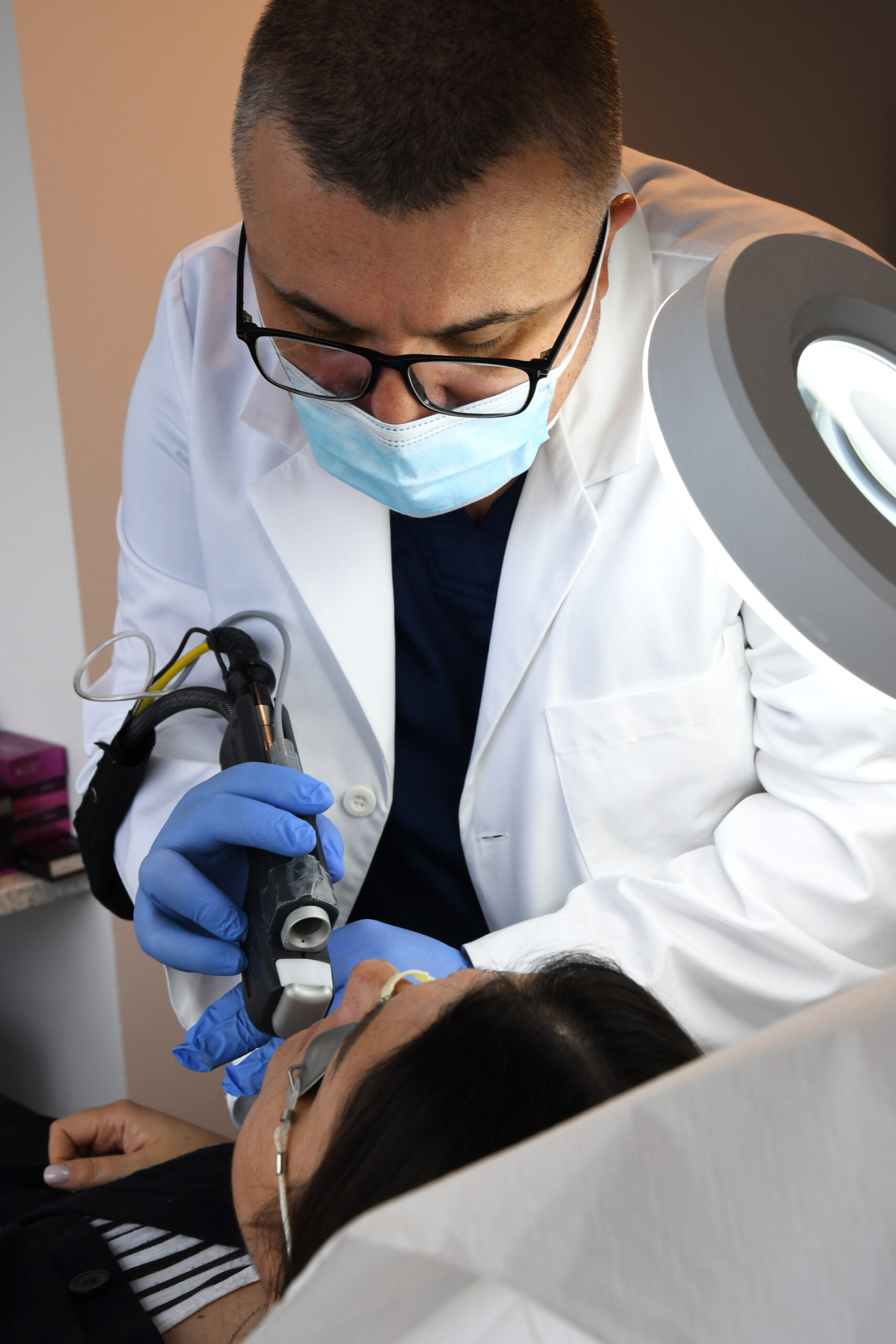
Fractional CO2 laser is successfully used in the treatment of stretch marks (striae distensae), by stimulating the regeneration of collagen and elastin. The procedure creates controlled microlesions in the skin, which favor the restoration of the affected dermal structure. The treatment can improve the texture and color of stretch marks, reducing their visibility. Multiple sessions are required for optimal results, and recovery is usually rapid.

Motivations and

Values